PET Scan: Complete Guide to Procedure, Uses, Preparation, Benefits and PET CT Scan
A PET Scan (Positron Emission Tomography) is an advanced medical imaging test that provides information about the metabolic activity of tissues and cells inside the body. Unlike imaging tests that mainly show the structure or anatomy of organs, a PET Scan can help doctors understand how certain tissues are functioning.
PET imaging has become an important part of modern diagnostic medicine, particularly in the evaluation and management of many types of cancer. It may also be used in selected neurological and cardiac conditions. In many cases, PET imaging is combined with CT imaging to create a PET CT Scan, providing both functional and anatomical information in a single examination.
For patients searching for a PET Scan in Chandigarh, understanding how PET imaging works, why it is recommended, how to prepare, what happens during the examination, and how the results are interpreted can make the process easier and more understandable.
What Is a PET Scan?
PET stands for Positron Emission Tomography. It is a nuclear medicine imaging technique that uses a small amount of a radioactive substance called a radiotracer to evaluate activity within the body.
The radiotracer is introduced into the body and travels to tissues according to its biological properties. The PET scanner detects signals from the tracer and creates images that show areas of different metabolic activity.
One of the most commonly used PET tracers in oncology is FDG (fluorodeoxyglucose), which behaves similarly to glucose. Because many cells use glucose for energy, FDG can help demonstrate differences in metabolic activity between tissues.
It is important to understand that increased uptake on a PET scan does not automatically mean cancer. Infection, inflammation, and certain normal tissues can also show increased activity. Therefore, PET findings must be interpreted in the context of the patient’s medical history, symptoms, laboratory tests, and other imaging studies.
What Is a PET CT Scan?
A PET CT Scan combines two imaging technologies:
PET – Positron Emission Tomography
Provides information about metabolic or functional activity.
CT – Computed Tomography
Provides detailed anatomical images showing the location and structure of organs and tissues.
By combining these technologies, doctors can identify areas of abnormal metabolic activity and understand their anatomical location more accurately.
For example, if a PET scan shows an area of increased activity, the CT component can help determine exactly where that area is located and what structural abnormality may be associated with it.
PET CT is particularly useful in many areas of oncology and is also used in selected other medical conditions.
Why Is a PET Scan Performed?
A PET Scan may be recommended for several clinical reasons. The exact indication depends on the patient’s diagnosis, symptoms, previous test results, and treatment plan.
One of the most common applications of PET CT is cancer evaluation.
Doctors may use PET CT imaging to:
- Evaluate certain suspected cancers
- Help characterize certain abnormalities
- Determine the extent or spread of some cancers
- Identify areas that may represent metastatic disease
- Assist in treatment planning
- Assess response to cancer treatment
- Monitor changes after treatment
- Investigate suspected recurrence in appropriate clinical situations
PET imaging can provide information that complements CT, MRI, ultrasound, biopsy, and laboratory investigations.
The decision to perform PET CT should always be based on the patient’s individual medical circumstances.
PET Scan for Cancer Evaluation
PET CT has an important role in modern cancer care.
Cancer cells can sometimes have different metabolic behavior compared with normal tissues. A PET scan using an appropriate tracer may identify areas with increased metabolic activity.
Depending on the type of cancer, PET CT can help doctors understand:
- Where the primary abnormality is located
- Whether nearby lymph nodes may be involved
- Whether disease may have spread to distant organs
- How extensive the disease appears to be
- Whether treatment is producing a response
- Whether an abnormal area requires further evaluation
PET CT findings can be particularly useful when combined with the patient’s clinical history and other investigations.
However, PET CT should not be considered a standalone test for every cancer. In many situations, tissue diagnosis through biopsy remains important for confirming cancer.
How Does a PET Scan Work?
The PET Scan process generally begins with patient preparation.
Depending on the type of PET examination, the patient may be instructed to fast for a specific period before the scan. Blood glucose levels may also need to be checked, particularly when an FDG-based PET scan is planned.
After arrival at the imaging facility, the healthcare team reviews the patient’s medical information and confirms the examination requirements.
A radiotracer is then administered, usually through an intravenous injection for FDG PET imaging.
The patient rests quietly while the tracer distributes throughout the body. This waiting period is important because it allows the tracer to reach the tissues that are being evaluated.
After the appropriate uptake period, the patient is positioned on the PET/CT scanner. CT images are acquired first or as part of the combined protocol, followed by PET imaging.
The patient needs to remain still during image acquisition.
PET Scan Preparation
Proper preparation can be important for obtaining good-quality PET images.
For many FDG PET scans, patients may be asked to fast for several hours before the examination. The exact fasting period should always be confirmed with the diagnostic centre.
Patients may also receive instructions regarding physical activity before the scan because strenuous exercise can affect glucose metabolism and potentially influence imaging.
Blood glucose levels may be checked before tracer administration.
Patients should inform the medical team if they:
- Have diabetes
- Take regular medications
- Are pregnant or may be pregnant
- Are breastfeeding
- Have recently undergone another medical procedure
- Have any relevant medical condition
- Have specific allergies or previous reactions to medications or contrast agents
Preparation requirements can vary depending on the type of PET scan and tracer being used.
Therefore, patients should follow the exact instructions provided by their healthcare professional or PET CT centre.
What Happens During a PET CT Scan?
On the day of the examination, the patient is generally asked to arrive at the centre according to the appointment instructions.
After registration and preparation, the radiotracer is administered.
The patient then rests in a designated area for a certain period while the tracer distributes throughout the body.
It is generally important to remain relaxed and avoid unnecessary physical activity during this period.
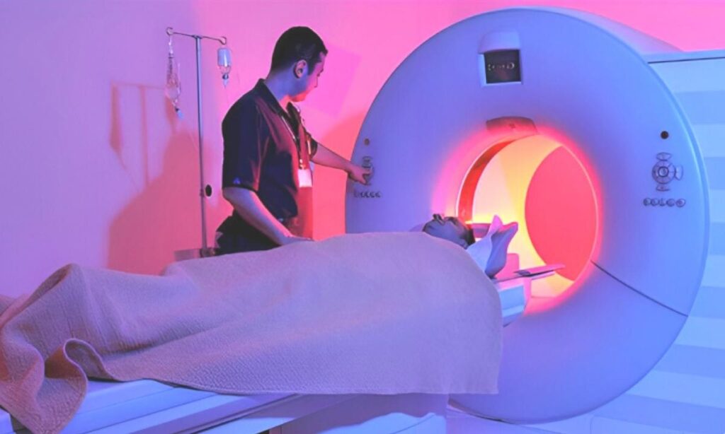
After the uptake period, the patient lies on the examination table of the PET CT scanner.
The table moves slowly through the scanner while the equipment collects PET and CT information.
The patient may be asked to remain completely still and follow breathing instructions when required.
The scan itself may take a variable amount of time depending on the examination protocol and the patient’s circumstances.
Once imaging is completed, the images are reviewed by qualified specialists.
Is a PET Scan Painful?
A PET Scan itself is generally not painful.
The main physical discomfort may be associated with the injection used to administer the radiotracer.
During the scan, the patient simply needs to lie still on the examination table.
Some patients may feel anxious about being inside the scanner. Informing the healthcare team about any concerns before the examination can help the staff provide appropriate guidance.
Is PET Scan Safe?
PET imaging uses a small amount of radioactive tracer. The tracer is selected according to the type of examination and is administered in a controlled medical setting.
The radioactive material naturally decreases and is eliminated from the body over time.
Healthcare professionals consider the expected diagnostic benefit when recommending a PET examination.
Patients should inform the medical team if they are pregnant, planning pregnancy, or breastfeeding. Special considerations may be necessary depending on the clinical circumstances and tracer being used.
Patients should always follow the post-scan instructions provided by the nuclear medicine or imaging team.
PET Scan and Radiation
Because PET uses a radioactive tracer and PET CT also includes CT imaging, the examination involves exposure to radiation.
The amount of radiation depends on several factors, including the tracer, CT protocol, scan type, and patient-specific requirements.
PET CT is recommended when the expected clinical value justifies the examination.
Modern imaging centres use established protocols designed to obtain clinically useful images while managing radiation exposure appropriately.
If you are concerned about radiation, discuss your concerns with your doctor or nuclear medicine specialist before the examination.
PET Scan vs CT Scan
PET Scan and CT Scan provide different types of information.
A CT Scan primarily provides detailed anatomical information. It can show the size, shape, and location of structures and abnormalities.
A PET Scan provides information about metabolic activity.
A PET CT Scan combines these two types of information.
This combination can be particularly valuable when doctors need to understand both the location and metabolic behavior of an abnormal area.
The choice between CT, PET, MRI, PET CT, or another imaging test depends on the patient’s condition and the clinical question that needs to be answered.
PET Scan vs MRI
MRI and PET CT are different imaging techniques.
MRI uses a magnetic field and radio waves to create detailed images, particularly of soft tissues. It can be extremely useful for evaluating the brain, spinal cord, joints, muscles, and various organs.
PET CT combines metabolic information from PET with anatomical information from CT.
In certain medical conditions, doctors may recommend MRI, PET CT, or both depending on what information is required.
Neither test is universally better than the other. The appropriate examination depends on the individual clinical situation.
What Does Increased PET Uptake Mean?
One of the most important aspects of PET imaging is understanding tracer uptake.
An area with increased tracer uptake may indicate increased metabolic activity. In oncology, this can sometimes correspond to active cancer.
However, increased uptake is not specific to cancer.
Inflammation, infection, healing tissues, and some normal physiological processes can also demonstrate increased uptake.
Therefore, PET findings should always be interpreted by a qualified specialist and correlated with CT findings, clinical history, previous imaging, laboratory tests, and other relevant information.
Understanding a PET CT Scan Report
After the examination, the PET and CT images are carefully reviewed by the appropriate specialist.
The final report may describe areas of normal and abnormal tracer uptake, anatomical findings, lymph nodes, organs, and other relevant observations.
The report may also compare current findings with previous scans when available.
Patients should discuss their report with the doctor who recommended the examination. A doctor can explain the findings in relation to the patient’s diagnosis and treatment plan.
It is not advisable to interpret isolated PET findings without considering the complete clinical picture.
PET CT for Treatment Monitoring
PET CT can sometimes be used to assess how a disease is responding to treatment.
For certain cancers, changes in metabolic activity may occur before obvious structural changes become apparent on conventional imaging.
Doctors may therefore use PET CT at appropriate stages of treatment to evaluate response.
The timing of follow-up PET CT is important and should be determined by the treating specialist because recent treatment, inflammation, or healing can influence tracer uptake.
PET CT and Cancer Staging
Cancer staging involves determining the extent of disease within the body.
Depending on the cancer type, PET CT can provide valuable information about the primary tumor, lymph nodes, and possible distant sites of disease.
This information may help the oncology team develop an appropriate treatment strategy.
However, the role and accuracy of PET CT vary between different cancers, so it is not appropriate for every cancer type or every patient.
PET Scan in Chandigarh
Patients looking for a PET Scan in Chandigarh may come from Chandigarh as well as nearby areas such as Mohali, Panchkula, Zirakpur, Kharar, and New Chandigarh.
When selecting a PET CT centre, patients should consider factors such as the availability of appropriate PET CT technology, qualified nuclear medicine and radiology professionals, trained technical staff, proper patient preparation, image quality, reporting expertise, and patient support.
Before booking a PET CT Scan, it is useful to confirm:
- The exact type of PET examination required
- Fasting instructions
- Blood glucose requirements, if applicable
- Medication-related instructions
- Expected appointment duration
- Whether previous reports are required
- Reporting process and expected turnaround time
Frequently Asked Questions About PET Scan
Is PET Scan only used for cancer?
No. Although PET imaging is widely used in oncology, it can also have applications in selected neurological and cardiac conditions. The appropriate use depends on the tracer, clinical question, and patient’s condition.
How long does a PET CT Scan take?
The total appointment time can vary because it includes preparation, tracer administration, the uptake period, imaging, and other necessary steps. Your PET CT centre can provide an estimated appointment duration.
Can I eat before a PET Scan?
For many FDG PET examinations, fasting is required for a specified period. However, preparation varies depending on the examination. Follow the instructions provided by your healthcare team.
Can diabetic patients undergo PET CT?
Diabetic patients can undergo PET CT when medically appropriate, but blood glucose management and medication instructions may require special planning. Diabetic patients should inform the PET CT team in advance.
Is PET CT the same as a CT Scan?
No. A CT Scan primarily provides anatomical information, while PET provides metabolic information. PET CT combines both technologies.
Does a PET Scan confirm cancer?
PET Scan findings alone do not always confirm cancer. Increased tracer uptake can occur in cancer as well as infection, inflammation, and some normal tissues. Doctors interpret PET findings along with other investigations.
Conclusion
A PET Scan is an advanced functional imaging technique that provides valuable information about metabolic activity within the body. When combined with CT imaging, PET CT Scan can provide both functional and anatomical information, making it an important tool in many areas of modern diagnostic medicine.
PET CT is particularly valuable in selected cancer-related situations, including disease evaluation, staging, treatment planning, treatment response assessment, and evaluation of suspected recurrence. Its role can also extend to selected neurological and cardiac conditions.
Proper preparation is an important part of the PET Scan process. Patients should carefully follow fasting, medication, blood glucose, and other instructions provided by their healthcare team.
If you have been advised to undergo a PET Scan or PET CT Scan in Chandigarh, discuss the examination with your treating doctor and follow the specific instructions provided by the imaging centre. The appropriate imaging test should always be selected according to the patient’s individual medical condition and clinical requirements.
Medical Disclaimer: This article is provided for general educational and informational purposes only. It does not replace professional medical advice, diagnosis, or treatment. PET Scan or PET CT Scan requirements, preparation, interpretation, and follow-up should be determined by qualified healthcare professionals based on the individual patient’s medical condition.